What is a stress echocardiogram?

A stress echocardiogram test, or “stress echo” for short, serves he same general purpose as a standard cardiac stress test. By observing how the heart behaves during exercise, the doctor performing or reading the test can get some clues about the kinds of problem that a person may or may not have.

The difference is that echocardiographic (ultrasound) images of the heart are obtained both at rest, before the exercise protocol begins, and immediately after exercise.  This gives additional information that can minimize some of the shortcomings of the standard stress test.

How is a stress echo conducted?

If you come for a stress echo, you will be asked to walk on a treadmill while connected to an ECG monitor.

The standard exercise protocol is the “Bruce protocol” which proceeds through stages, very easy at first, and gradually becoming more difficult. The initial stage is a low walk at a 10% incline.  With each successive stage the incline increases and the speed also increases gradually.  This allows you to warm up, to get used to the equipment and to relax before the effort gets more intense.  It also gives a chance to the doctor and the technician to observe the heart response more deliberately. Because the stress test starts off nice and easy, it provides a good opportunity to chat, exchange jokes, and get to know one another!

Once an adequate effort level is achieved, you will be asked to lie down on the examination table where images will be obtained while the heart is still pumping fast.  Any abnormal motion of the heart or abnormal pressure is noted at that point.  You can then rest for a few minutes while the ECG machine continues to record your heartbeat.

Must we fear the Apple Watch’s new ECG feature?

UPDATE March 20, 2019:  The results of the “Apple Heart Study” were just released at the annual meeting of the American College of Cardiology.  Read my comment at the end of this post for an update.


Today, Apple released its ECG app and irregular heart rhythm notification feature.

The announcement about the technology was made with great fanfare a couple of months ago, when Apple also revealed that the device had received approval by the FDA.  This raised some eyebrows because—according to some—the technology was being approved and released to the public without evidence of effectiveness or harm.

What is so unique and novel about the product is that it claims to be able to detect atrial fibrillation, or “a.fib,” which is a common irregularity of the heart rhythm.  A.fib is not usually fatal but it can be a problem for two reasons.

First, the arrhythmia causes and irregularity and rapidity of the pulse that can provoke palpitations and also impair the pumping capacity of the heart.  This typically causes symptoms, so that people who develop a.fib typically become aware of the problem and seek medical attention relatively promptly.

But not everybody is necessarily aware that they have a.fib.Read more

Great news about the coronary calcium scan

As readers of the blog may know, I have been a very strong proponent of the coronary calcium scan for many years.  The scan is a great tool to help determine vascular health and make a decision regarding the treatment of high cholesterol.

I have argued in previous blog posts and videos that patients with a coronary calcium score of zero may be able to avoid medications to lower cholesterol.  In the last couple weeks, 2 news items from the cardiology community have bolstered my opinion.Read more

The ambulatory blood pressure monitor

The blood pressure is fickle

We typically think of the blood pressure as a pair of numbers that are measured in the doctor’s office once in a while.  If the numbers are persistently too high, you have “high blood pressure” (also known as hypertension) and that’s a concern for the future.  If untreated, hypertension can lead to serious complications, including heart, kidney and brain damage.

But we know that people can get a little nervous when they’re at the doctor’s office, so the blood pressure may be high there but be normal the rest of the time.  That is called “white coat hypertension,” because the stress of seeing a doctor in a white coat is enough to raise the blood pressure!

And we also know that the opposite can occur: the blood pressure may be deceptively low at he doctor’s office, but much higher during the day when the stress of work or of taking care of the household is in full force.  That is called “masked hypertension,” because the doctor only sees normal blood pressure numbers in the clinic and is unaware that the numbers are actually higher most of the time.

The blood pressure changes all the time, so it’s quite challenging to make a determination about hypertension on a few sets of numbers taken here and there.

A new way to measure BP

A few years ago, medical device manufacturers developed ambulatory blood pressure monitors (ABPMs), which are simple blood pressure machines, the size of a WalkMan tape player (does anyone remember those?!).

The device can be worn over a period of 24 to 48 hours and which automatically obtain a large number of measurements while you’re out and about during the day, and even at night when you sleep!Read more

Getting intimate with the carotid intima

We are pleased to announce the addition of the “carotid intima-media thickness” test (CIMT) to the array of non-invasive diagnostic tools available at Athletic Heart SF.

The idea behind the CIMT is simple: an ultrasound image of the carotid artery can provide information about the health of the artery itself and, more generally, about the overall health of the cardiovascular system.

The ultrasound image can identify two conditions.  First, the ultrasound can detect if there is actual plaque build-up which, if advanced, can block the flow of blood and contribute to the risk of stroke.  Fortunately, advanced plaque build-up is relatively rare.

Second, even in the absence of plaque build-up, the thickness of the vessel wall (CIMT) can give a broad indication of cardiovascular health.  The intima is the name of the inner lining of a vessel wall, and the media is the middle layer.  Both are seen in the image below as the bright thin line and the thin darker band behind it.

Ultrasound image of a carotid artery. The thin intima-media layer is indicated by the arrow.

Read more

When endurance athletes have “hearts of stone”


I have just returned from attending a course on the “Prevention of Sudden Cardiac Death in Athletes,” hosted by the University of Washington medical school in Seattle.  It was a terrific conference at which academic leaders in the field gave updates on the latest research.

Dr. Aaron Baggish, from the Massachusetts General Hospital in Boston, summarized two recent studies that have persuasively shown that coronary calcifications are more common in long-term endurance athletes compared to more sedentary controls.

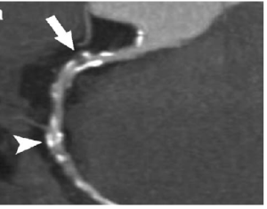
When the studies were published this past summer, Dr. Baggish was asked to write an editorial commentary to share his perspective.  That editorial was provocatively subtitled “Hearts of Stone.”  The phrase refers to the appearance on CT scans of hearts with heavily calcified arteries as shown in this image:

Calcium in coronary artery detected by CT scan.
Image attribution: Wikimedia Commons

Background

In the last several years, a few reports were published suggesting that endurance athletes may be more prone to having build-up of plaque and calcium in their coronary arteries.  Those reports presented a paradox, because we also know beyond any doubt that regular, moderate level exercise promotes cardiovascular health and longevity.  The studies raised the concern that exercise could be harmful after a certain point.Read more

Are statin drugs over-prescribed?

Your heart health in a heartbeat – Episode 1

If your cholesterol is high, should you immediately start lifelong medication?  Join me as I discuss a new study which addresses this important question.

Link to the study discussed in this episode:

Implications of Coronary Artery Calcium Testing Among Statin Candidates According to American College of Cardiology/American Heart Association Cholesterol Management Guidelines

Related blog post about the heart scan:

The heart scan as a “mammogram of the heart”


The echocardiogram: seeing with sounds!


Ultrasound technology is not exactly new, but it never ceases to amaze me.

First developed in the 1950’s, medical ultrasound technology uses the fact that different tissues and fluids in the body reflect sound waves differently.  If an ultrasound source is applied over the chest, the sound will be reflected back from different parts of the heart in a way that can be analyzed to reconstruct an image of the heart in real time.

"Ventricular Septal Defect". Licensed under Public Domain via Wikimedia Commons.

Licensed under Public Domain via Wikimedia Commons.

Because of the clarity and reliability of the image produced, echocardiography has become an indispensable tool of cardiac diagnosis and has allowed doctors to “see” directly what they previously had to imagine, deduce, or guess: the size of the heart chambers, the pumping strength of the heart muscle, the health and function of the heart valves, the presence of fluid build up around the heart, the size of the aorta and other major vessels, and many other important cardiovascular features.  Having all this information at the tip of our fingers would have been unimaginable to cardiologists of a generation or two ago.

What’s more, the procedure itself—as far as the patient is concerned—is completely safe and painless.  The technician applies some gel on the chest, which allows the ultrasound probe to have better contact with the skin.  The probe itself is like a speaker and microphone in one piece, transmitting and receiving the sound waves which are immediately analyzed and displayed on a monitor.

Of course, there is a great deal of skill involved in obtaining good images.  Because the heart is surrounded by ribs and lung tissue which interfere with the ultrasound images of the heart, one must find adequate “windows” to take a peak at the heart.

The procedure for a complete echocardiogram takes about 30 minutes or so.

Introducing the Coronary Health Starter Kit!


Dear Friends,

In an effort to make heart scans more widely available, I am delighted to introduce a new service:  AHSF’s Coronary Health Starter Kit.

This service consists of an affordable telephone consultation focused on a cardiac prevention.  As part of this service, I order the famed “mammogram of the heart“, the coronary heart scan that can detect the presence of coronary artery plaques in their early stage, before they cause complications such as heart attacks and sudden cardiac arrest.

I recommend this service for men over age 40 and for women over age 50 (or younger women  if they had premature menopause).  You may consider obtaining the service at an earlier age if you have strong risk factors, such as a family history of coronary disease at a young age. This is a screening service, so it assumes that you feel generally well and have no specific symptoms to report.

I cannot think of a better investment for your heart health than to take advantage of this service and obtain a heart scan.  Please do not delay.  It is as simple as picking up the phone and calling 1-415-567-1014 between 9a-3p to make time for a telephone consultation.Read more

The “mammogram of the heart”


The phrase “mammogram of the heart” refers to the heart scan that detects calcium build-up in the coronary arteries.  The phrase was coined by the pioneers in this technology to try to impress on the public and on the medical community the simplicity and value of this test.

Calcium in coronary artery detected by CT scan.Image attribution: Wikimedia Commons

Calcium in coronary artery detected by CT scan.
Image attribution: Wikimedia Commons


Just as a mammogram can identify cancer before it spreads, a heart scan can identify the presence of coronary artery disease before it causes symptoms.  And like the mammogram, the heart scan is easy to perform, uses a low amount of radiation, and is inexpensive.Read more